10 Months of deep sky astrophotography

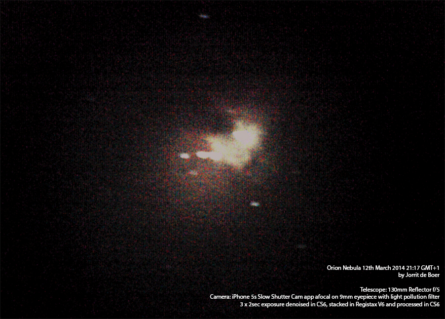
When I started this site, I just began taking my first astrophotos. The image below was my first attempt to capture the Orion Nebula with my iPhone.

First try at m42 with iPhone

First try at m42 with iPhone

After this first attempt, I improved my astrophoto skills by practicing with planetary imaging. But still, I couldn’t squeeze any more out of the iPhone than this:

Final try at m42 with iPhone

Final try at m42 with iPhone

I decided it had been my final try at iPhone deep sky imaging. I tried my luck with both xbox live and ps3 webcams, but the xbox live cam didn’t pick up anything special and I still haven’t got the ps3 cam to do long exposures.

After I bought my DSLR I could do untracked 0.8 second exposures without too much trailing, but the 0.8 second exposures aren’t that exciting really. It’s my new motorized tracking mount in combination with my DSLR that makes all the difference. When last week the thick layer of clouds showed the tiniest opening, I immediately set up my telescope to take advantage. Unfortunately the opening was so small, that by the time I had everything up and running – which didn’t take that long – a milky but slightly transparent blanket covered the skies again. To make things worse, some serious winds shook my telescope tubes every now and then.

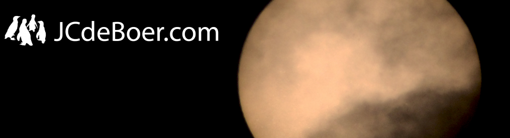
Bright as the Moon is, I could just lower the ISO values and take a decent picture of it through the clouds. To give you an idea of the amount of clouds present, I’ve also added a few images taken at higher ISO values so that you can see the clouds passing in front of the Moon.

Moon at low ISO values

Moon at low ISO values

Despite the once more disappointing skies and the feisty gusts of wind, I decided to try finding the Orion Nebula. After a while I succeeded in doing so and started taking 20 second exposures between successive wind gusts and through the clouds. The majority of the images showed massive trailing and were useless. Some however, were not that bad and after 30 minutes or so I had 4 or 5 decent shots.

After stacking and some post processing, I was suprised to find that I had also captured the Running Man Nebula, which isn’t as bright as the Orion Nebula itself. All in all, the small stack of photos, taken at seriously bad conditions, ended up in my best photo of the Orion Nebula yet. I can’t wait to test my setup under really good conditions 🙂

M42 taken with DSLR and tracking mount through a thin layer of clouds

M42 taken with DSLR and tracking mount through a thin layer of clouds

Microscopes do not care about clouds

Except for 2 evenings, my motorized telescope has been collecting dust now for about 2 months. And on those 2 evenings, seeing became poor around midnight. I really can’t remember another period this worse for amateur astronomers. Luckily though, I don’t need clear skies to use my microscope.

I have a few (microscope) images from last month that I haven’t shown you yet and also some more recent photos.

Starting with a few microscopic images of elemental gallium. Gallium is fun to play with because it melts at 30 degrees Celcius (i.e. in your hand) and it isn’t as dangerous as mercury. Actually, it’s not harmful at all as long as you don’t eat it. And the best thing is, I just happen to have some at home for entertainment purposes.

For the images I deposited some droplets of liquid gallium on my glass slide and allowed it to crystallize slowly. The resulting lumbs of solid gallium were somewhat edgy but also contained plenty of rounded corners as you can see in the image below.

Small crystallized droplet of pure gallium.

Small crystallized droplet of elemental gallium at 40x.

I was really fascinated by this image. It seems to be bell-shaped and contains razor sharp cornered edges around curved bodies. This image was taken at 40x magnification. Someone suggested that the straight edges may be due to expansion of the material after the outer shell had solidified.

The next image also shows some very interesting feautures. Very apparent are the colors in the top left. I believe this is due to the use of both achromatic objectives and poor lighting. The microscope I have is fitted with a bulb underneath the sample and as you may expect, light isn’t really visible through lumbs of metal. So I had to use a lamp that I positioned so that it illuminates the sample from the side.

On the right side of the image, the flat surface isn’t very flat. It shows a funny structure that may be due to the fact that the gallium crystallized under water and didn’t have the chance to dry properly.

Another crystallized gallium droplet

Another crystallized gallium droplet

I shot the following two images during a beautiful november sunset. On the first you can se a very faint second rainbow above the brighter one. The second image shows how magnificently golden the sky was. Normally I like to use some photoshop to subtly enhance the best features of an image, but I didn’t really edit this one. The photo was just perfect.

A quite faint double rainbow

A quite faint double rainbow

Golden evening skies over Enschede

Golden evening skies over Enschede

Do you remember this image I shot of a begonia? I used a telescope to take it.

Begonia through a 900mm telescope

Begonia through a 900mm telescope

Well, here is the same begonia through a microscope:

Red leaf of a begonia through the microscope

Red leaf of a begonia through the microscope at 40x

I really love it’s brilliant red color. The image below shows the same area but at higher magnification. Stacking the differently focused images was quite a hastle, but it worked out okay for these images. I had one at even higher magnification (400x), but stacking this produced a blur.

The same red leaf at higher magnification

The same red leaf at 100x magnification

Nylon festival wrist band trough the microscope

Nylon festival wrist band trough the microscope

The image above shows a close up at 100x of a nylon wrist band I got from a music festival. Purely synthetic. Absolutely not synthetic, is the waterweed in the image below. It shows all the little green chloroplasts that give the plant its green color. The way you see it here is somewhat different from what I saw through the microscope. I could see a lot of cytoplasmic (the liquid that fills the volume of the cell) streams, which is highlighted by lots of chloroplasts that travel through the leaf. Maybe I will make a video of that some day 🙂 .

Waterweeds trough the microscope

Waterweeds trough the microscope at 100x